Chapter 6: Infections and Infestations
Infectious Exanthems
An exanthem is a widespread rash that can be triggered by an infection as well as other causes such as medications. Infectious exanthems are especially common in children and may have characteristic features depending on the causative organism.
Non-Specific Viral Exanthem
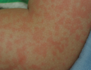
Non-specific viral exanthems are the most common exanthems in children. They present as red macules and papules that are blanchable (redness fades when pressure is applied), distributed widely on the trunk and extremities, and often coalesce. The rash is often associated with viral symptoms such as fever and might be difficult to distinguish from a morbilliform drug eruption. There are numerous viruses which cause non-specific viral exanthems including enterovirus, adenovirus, parainfluenza and respiratory syncytial virus.
Erythema Infectiosum
Erythema infectiosum, also known as “fifth disease”, is caused by infection with Parvovirus B19. This is a common disease of school-aged children and typically occurs during the winter and spring. The exanthem occurs approximately 1-2 days following a prodrome of mild fever and headache. It begins as a distinct “slapped cheek” appearance with bright red patches to both cheeks. This is typically followed by a lacy red rash on the extremities that lasts for 1-3 weeks. There is no specific treatment and affected children can attend school, as the infectious stage occurs before the rash is evident.
Measles
With high vaccination rates in many countries, measles is becoming a less common disease worldwide. Still, it is a cause of significant morbidity and mortality globally and is highly contagious with up to 90% of susceptible people who get exposed contracting the disease. Outbreaks continue to occur even in countries with high vaccination rates and have been seen frequently in recent years, especially in populations with high rates of vaccine avoidance. Measles is caused by a single-stranded RNA paramyxovirus. It is transmitted by air-borne droplets from 1-2 days before the onset of symptoms until 3-4 days after the rash appears. Patients experience a prodrome of cough, coryza (runny nose), and conjunctivitis. The first skin lesions are called Koplik spots and are 1-2 mm blue-white macules on the oral mucosa (typically the inner cheeks). The rash appears about 2 weeks after exposure and 2-4 days after the beginning of symptoms. It is characterized by non-pruritic macules and papules beginning on the head and neck then spreading to the trunk and extremities (cephalocaudad spread). Treatment is with supportive care, vaccination of any unvaccinated contacts, and Vitamin A supplementation in children who contract the disease. This supplementation has been shown to decrease mortality by 30% in children and works to strengthen the mucosal barrier in the respiratory and gastrointestinal tracts. Complications can be serious and include pneumonia, encephalitis and myocarditis.
Rubella
As with measles, the incidence of rubella has decreased significantly with the advent of routine vaccination. As a result, it is very uncommon in most of the world; however, it is still of clinical importance due to some of its serious complications and the risk of fetal infection which can cause significant congenital abnormalities. A prodrome of fever, headache and malaise is followed 5 days later by an exanthem of “rose-pink” macules that starts at the head and travels downward (cephalocaudad spread). There may also be small red dots on the soft palate accompanying the exanthem which are known as Forcheimer spots. In most healthy children and adults, the disease is self-limiting and treatment is supportive.
Scarlet Fever
Scarlet fever is a bacterial illness due to toxins produced by Streptococcus pyogenes and was often fatal in the pre-antibiotic era. It most often occurs in children aged 4-8 and is associated with streptococcal pharyngitis (“strep throat”) or impetigo (superficial skin infection with S. pyogenes). It typically begins with fever, sore throat and swollen neck glands with a distinct exanthem appearing 12-48 hours following this. The exanthem consists of tiny pink to red spots that cover most of the body and have a characteristic “sand paper” texture. The tongue is often swollen and red (“strawberry tongue”). Diagnosis can be assisted with a throat swab showing growth of S. pyogenes or with anti-streptolysin-O titres. The treatment of choice is penicillin for 10-14 days – a complete course is important to reduce the risk of complications such as rheumatic fever and post-streptococcal glomerulonephritis.
Gianotti-Crosti Syndrome
Gianotti-Crosti syndrome was initially associated with Hepatitis B infections but more recently has been shown in association with various other viral infections (EBV, CMV, adenovirus, etc.) and some non-viral infections (S. pyogenes, Mycoplasma pneumonia). It is seen in children aged 6 months-14 years and causes monomorphous (all the lesions have a similar appearance), flat-topped, pink/brown, edematous (swollen) papules most often located on the knees and elbows and less often on the face and buttocks. The trunk is typically spared. Lesions last for over 10 days, but do not require any treatment and resolve spontaneously. Rarely, the rash can last up to 8 weeks.
Kawasaki Disease
Kawasaki disease is a multisystem disease that generally affects children less than 5 years old. While the exact cause still remains unknown, infectious etiologies have been postulated. Kawasaki disease is significant for being the number one cause of acquired heart disease among children in North America. The classic findings include fever lasting more than 5 days, redness of the conjunctivae, unilateral cervical lymphadenopathy, swelling/redness of the hands/feet, and “strawberry tongue”. An exanthem is present in ~80% of cases but its appearance is variable. Most commonly it is widespread red macules and papules similar to measles, and may favor the perineal area. If caught early enough the treatment of choice is intravenous immunoglobulin (IVIg). Patients should be referred to cardiology to assess for any cardiac involvement.
