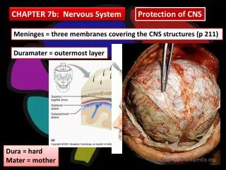
Download
1 / 103
1.09k likes | 1.39k Views
Chapter 10 Physiology of Nervous System. Section 1. The Functions of Neuron and Neuroglia. Neuron. The elementary functions of neuron Receive the excitations or inhibitions induced by internal or external stimulations. Analyze and integrate the information from every organs.

E N D
Section 1 The Functions of Neuron and Neuroglia
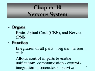
Neuron Theelementary functionsof neuron • Receive the excitations or inhibitions induced by internal or external stimulations. • Analyze and integrate the information from every organs. • Generate or carry the demands regulating the activities of the effectors. • Some neurons have neuroendocrine function.
Neurons have 4 important zones • Soma and dendrites–receive the information,generate and integrate the local potential changes. • Initialsegment- action potentials are generated. • Axonprocess- transmits the impulses to the nerve endings. • Nerveendings- release the synaptic transmitters.
Axoplasmic transport • Anterograde anxoplasmic transport: soma → terminals -Rapid transport : 410mm/d, organelles with membrane, neurotransmitters( neuropeptide), mitochondria and enzymes - Slow transport: 1-12(0.5-10)mm/d. microtubuleand microfilament, • Retrograde axoplasmic transport : soma ← terminals 205mm/d. NGF, virus and toxin,etc. by endocytosis.
Neurotrophin • Nerve growth factor(NGF) • Brain-derived neurotrophin factor(BDNF) • Neurotrophin 3 • Neurotrophin 4/5 • Neurotrophin 6 • Ciliary neurotrophin factor(CNTF) • Glial cell-derived neurotrophin factor(GDNF) • Leukemia inhibitory factor(LIF) • Insulin-like growth factorⅠ(IGF-Ⅰ) • Transforming growth factor(TGF) • Fibroblast growth factor(TGF) • Platelet-derived growth factor(PDGF)
Neuroglia • About 1.0×10¹²~ 5.0×10¹² neuroglia cells , 10~50 fold of neurons • Dendrites and axons can not be distinguished clearly • No synapse formed and no AP produced
The types of glia • CNS - astrocyte oligodendrocyte microglia ependamal cell Choroidal epithelium • PNS - Schwann cell satellite cell
Functions of glial cells • Astrocytes (Astroglia) -Support the neurons - Clean up brain "debris"( damaged material) and fill in the damaged area - Transport nutrients to neurons - regulate the external chemical environment of neurons by removing excess ions, and recycling neurotransmitters.
Transport nutrients to neurons, • They regulate the external chemical environment of neurons • by removing excess ions, notably potassium, and recycling • neurotransmitters released during synaptic transmission
Oligodendrocytes and Schwann cells -myelinate axons (1) insulate the axons (2) facilitate the conduction of electrical impulses.
Microglia -act as the immune cells of the CNS -remove most of the waste and cellular debris from the CNS -derivation,action in brain injury, action in other diseases.
Section 2 General interactions between neurons
Classificationsof Synapses – Chemical synapses ▪Directed synapses (Typical synapses) ▪Non- directed synapses (Varicosity) – Electrical synapses
TypicalSynapses (chemical synapses) Synapse The small gap or space between the axon terminals of one neuron and the dendrites or cell body of the next neuron is called the Synapse .
Structure of Synapse • Membrane of presynaptic neuron • Synaptic cleft • Membrane of postsynaptic neuron
MajortypesofSynapses A: axo-somatic synapse B:axo-dendritic synapse C:axo-axonic synapse
Processof Typical Synaptic Transmission 1. An arrivingaction potentialdepolarizes the presynaptic membrane. 2. Calcium ionsenter the cytoplasma of the synaptic knob. 3. Neurotransmitters release. 4. Neurotransmittersdiffuseto andbindto thereceptorson postsynaptic membrane. 5. Receptorson the postsynaptic membrane are activated, producing apostsynaptic potential. 6. Neurotransmitters arebroken down.
Electrical Activities of Postsynaptic Neurons (Postsynaptic Potential) Forms of the postsynaptic potential • Excitatory postsynaptic potential (EPSP) • Inhibitory postsynaptic potential (IPSP)
PostsynapticPotentials Excitatory postsynaptic potential • When a neuron responds to the neurotransmitter postsynaptically, it allows ions to move across its membrane. • The movement of ions changes the membrane potential of the postsynaptic neuron. • It is called the “postsynaptic potential”. Inhibitory postsynaptic potential
EPSP • Excitatory transmitters → Synaptic cleft → bind to receptors → ↑the postsynaptic membrane's permeability to Na+, Ca2+ → enter the postsynaptic neuron →produce a depolarizing potential.
IPSP • Inhibitatory transmitters→ Synaptic cleft → bind to receptors → the postsynaptic membrane’s permeability toCl-(or K+) →Cl-enter the postsynaptic neuron →generate ahyperpolarizing potential. They can also produced by closure of Na+ or Ca2+channels
The EPSP is produced by depolarization of the postsynaptic membrane. During this potential, the excitability of the neuron to other stimuli is increased, and this the potential is called the EPSP. • The IPSP is produced by hyperpolarization of the postsynaptic membrane. During this potential, the excitability of the neuron to other stimuli is decreased, and this the potential is called the IPSP.
Types of postsynaptic potentials EPSP: • excitatory postsynaptic potential • can help lead to the production of an action potential • causes a depolarization IPSP: • inhibitory postsynaptic potential • can help to prevent the production of an action potential • causes a hyperpolarization
Features of post-synaptic potential • Post-synaptic body works as a “ device of integration ”. • Algebraic sumof EPSP and IPSP generated simultaneously. summation
InactivationofNeurotransmitters 1. Be reuptaken by presynaptic membrane or by Glial cells (Serotonin,NE) 2. Diffusion (Neuropeptide) 3. Enzymatic degradation (ACh )
Synaptic inhibition It can be divided into the postsynapticinhibition and presynapticinhibition according to the location.
Postsynapticinhibition The inhibitory interneuron releases the inhibitory neurotransmitter which induces the postsynaptic membrane hyperpolarizing. • Afferent collateral inhibition:The branches of axon synapse the inhibitory interneuron, which connect with other excitatory neurons. (reciprocal inhibition,交互抑制) • Recurrentinhibition:The inhibitory interneuron is connected in such a way that they act back on the excited cell itself.
Afferent collateral inhibition It is very important for coordinating reflex activity.
Strychnine(士的宁): block the inhibitory synapses glycin Recurrent inhibition
Presynapticinhibition • In presynaptic inhibition, there is no change in the postsynapstic membrane, but a reduction in the release of transmitter at the presynaptic terminal of the excitatory synapses. • Presynaptic inhibition is induced by an activation of axo-axonic synapse (combined with axo-somatic synapse).
A C C
Characteristicsof chemical synaptic transmission 1. One way conduction 2. Synaptic delay:0.5ms↑ 3. Summation 4. Susceptibility:to asphyxia, ischemia, and drugs. 5. Fatigue
Non-synaptic chemical transmission (Varicosity) • The autonomic nerve fibers • Smooth muscle cells • Distinguish it with neuromuscular junctions
Electrical synaptic transmission --- gap junction • Gap junctions allow charges to flow from on cell to the next. These ions may depolarize the adjacent cell to threshold. An AP is generated.
Section 3 Nerve reflex and neuronal circuit
neuronal circuit Divergence:responsible for motor and sensory systems. Convergence: responsible for the interpretation of the sensory stimuli. Chain circuit: facilitate or inhibit the signals. Recurrent circuit:prolong or shorten the signal activities.
Recurrent circuit Chaincircuit
Section 4 Sensory Function of Nervous System
Sensory pathways of somatic sensory gracile (薄束) and the cuneate Proprioception, active tactile Pain and temperature
Thalamus • The somatosensory thalamus is a relay station for most sensory modalities. • Axons from every sensory system (except olfaction) synapse here as the last relay site before the information reaches the cerebral cortex. • There are other thalamic nuclei that receive input from cerebellar-, basal ganglia- and limbic-related brain regions.
Thalamic relay nuclei • Sense-relay nuclei Relay auditory, visual and somatosensory impulse to the auditory, visual cortex and postcentral gyrus. • Associated nuclei which are concerned with the integration of movement, and the connection of the all senses. • The third thalamic nuclei They transmits nerve impulses to subcortical areas before transmission to the cerebral cortex. And then project diffusely to the cortex and maintain wakefulness.
Sensory Projection System • Specific projection system -From the Sense-relay nuclei and Associated nuclei -Projecting to the specific areas of cortex( point to point) - Few synapses only - Inducing the specific sensory. • Non-Specific projection system - From the third thalamic nuclei - Projecting diffusely to the cortex - Multisynapses - Maintaining the awareness state (ascending reticular system)
Ascending reticular activating system • Exerts an activating influence on the cortex to keep awaken state. • On the background of non-specific sensory pathway. • Poly-synaptic relay system.
Somatic Sensory areas in cerebral cortex • Somatic representations Somatic sensory area Ⅰ–postcentral gyrus Somatic sensory area II– between precentral gyrusand insula • proprioceptive representations - precentral gyrus -(sharing the same area with motor)